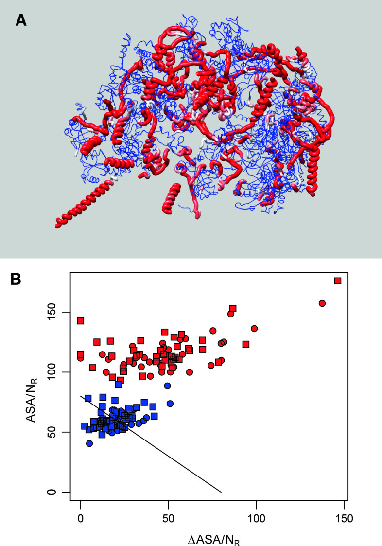
Fig. 2.
Foldability of globular and extended domains of ribosomal proteins from the yeast Saccharomyces cerevisiae. a Worm representation of 60S proteins. Color and width of ribbon corresponds to the OC posterior probability, where regions with a high probability are red and wide and regions with a low probability are blue and thin. b Nussinov’s plot of ΔASA against the ASA for the IC (blue) and OC (red) residues of 40S (circles) and 50S (squares) proteins

