Fig. 1.
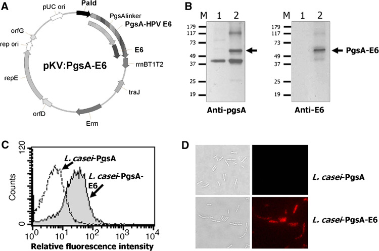
Surface expression of HPV16 E6 on L. casei. a A schematic diagram of the pKV-HPV16E6 plasmid used for HPV16 E6 expression on the surface of L. casei. b E6 protein expression in L. casei, as demonstrated by Western blot analysis using anti-pgsA3D (left) and anti-HPV16 E6 (right) polyclonal antibodies. M molecular weight markers, lane 1 L. casei-PgsA, lane 2 L. casei-PgsA-E6. A ~61-kDa protein band corresponding to the expected size of the PgsA-E6 fusion protein is present in lane 2. c FACS histogram of L. casei-PgsA (open) and L. casei-PgsA-E6 (filled) cells probed with polyclonal anti-HPV16 E6 antibodies, followed by an Alexa Fluor 594-conjugated anti-goat IgG antibody. d Representative immunofluorescence images of L. casei-PgsA and L. casei-PgsA-E6 (right) and corresponding differential interference contrast images (DIC) (left) are shown (×1,000)

