
Fig. 8.
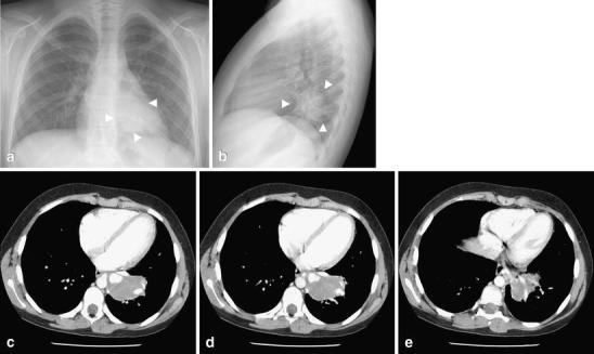
Intralobar sequestration in a 12-year-old girl with chronic cough. a Frontal and (b) lateral chest radiographs show a round opacity (arrowheads) with well-defined margins in the left lower lobe. c–e Contrast-enhanced CT demonstrates a round soft-tissue mass (arrows) in the posteromedial left lower lobe without tracheobronchial airway communication, but with arterial supply by a branch from the descending thoracic aorta and a draining pulmonary vein into the left atrium. The age of the girl and the symptoms should alert the radiologist about a possible underlying lesion. Compare these images with those in Fig. 2 (round pneumonia)