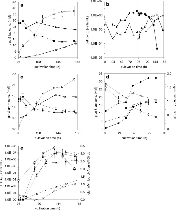
Fig 5.

Release and uptake of metabolites, cell concentrations, and virus titers during growth of MDCK and Vero cells on Cytodex 1 microcarriers (2 g/L) in SC/ΔSC medium in a 5 L STR (0–96 h) and subsequent influenza A/Wisconsin/67/2005 HGR H3N2 virus production (cell line adapted viruses) (96–168 h) with washing steps and medium exchange (ΔSC medium) (details see Table 1): MDCK cells (squares) compared to Vero cells (triangles). a Glucose (full symbols), lactate (empty symbols), b cell concentrations: cells on MC (full symbols), cells in suspension (empty symbols), c glutamine (full symbols), ammonia (empty symbols); d metabolite profiles for cell growth of Vero cells only (data for MDCK cells were as in (Genzel et al. 2004): glucose (white square), lactate (black square), glutamine (white circle), ammonia (black circle), glutamate (✱); e) glutamate (grey symbols), virus titer in log10 HA units/100 μL (full symbols), infectious virus titer (TCID50) (empty symbols). Relative standard deviations were as given previously ((Lohr et al. 2009)); read-out for the more sensitive HA assay: log10 HA units/100 μL with a relative standard deviation of 9.3%
