
Fig. 2.
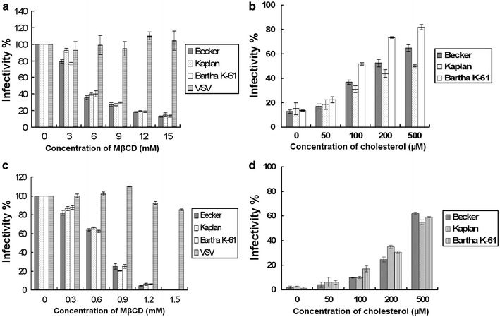
Importance of cholesterol on virus infection. Cells were treated with MβCD concentration range from 0 to 15 mM, and then cells were infected with three strains of PrV or VSV. The virus infection efficiency is shown in a. Vero cells treated with 12 mM MβCD were replenished with 50–500 μM exogenous cholesterol and then were subjected to PrV infection. The recovery of the viral infectivity is shown in b. The 100% infectivity values of PrV and VSV represent average plaque numbers of 300 and 150, respectively. Results are mean values of three independent experiments with standard deviation. PrV and VSV were treated with MβCD range from 0 to 1.5 mM, and the MβCD-treated viruses were used to infect cells. The infectivity of the viruses is provided in c. The PrV particles were treated with MβCD at the concentration of 1.2 mM and replenished with the exogenous cholesterol at the concentration ranging from 50 to 500 μM. d After replenishment, the infectivity of the viruses is determined. The 100% infectivity values of PrV and VSV represent average plaque numbers of 300 and 150, respectively