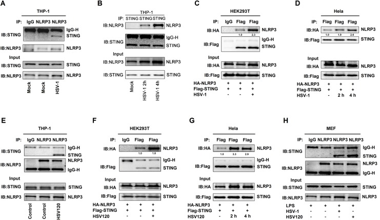
Fig 2. HSV-1 infection promotes the STING-NLRP3 interaction.
(A, B) TPA-differentiated THP-1 macrophages were mock-infected or infected with HSV-1 (MOI = 1) for 4 h (A) or 2 h and 4 h (B). (C, D) HEK293T cells (C) and Hela cells (D) were co-transfected with pFlag-STING and pHA-NLRP3, and then infected with HSV-1 (MOI = 1) for 2–4 h. (E) TPA-differentiated THP-1 macrophages were transfected with HSV120 (3 μg/ml) by Lipo2000 for 4 h. The Lipo2000 was used as the control. (F, G) HEK293T cells (F) and Hela cells (G) were co-transfected with pFlag-STING and pHA-NLRP3 and then transfected with HSV120 (3 μg/ml) for 4 h. (H) Primary MEFs were primed with LPS (1 μg/ml) for 6 h, and then infected with HSV-1 (MOI = 1) for 4 h or transfected with HSV120 (3 μg/ml) for 4 h. (A–H) Cell lysates were subjected to Co-IP using IgG (Mouse) or anti-NLRP3 antibody (A), anti-STING antibody (B), IgG (Mouse) or anti-Flag antibody (C), anti-Flag antibody (D), IgG (Mouse) or anti-NLRP3 antibody (E), IgG (Mouse) or anti-Flag antibody (F), anti-Flag antibody (G), and IgG (Mouse) or anti-NLRP3 antibody (H), and then analyzed by immunoblotting using anti-NLRP3 and anti-STING antibody (top) or analyzed directly by immunoblotting using anti-NLRP3 and anti-STING antibody (as input) (bottom). Densitometry of the blots were measured by Image J.

