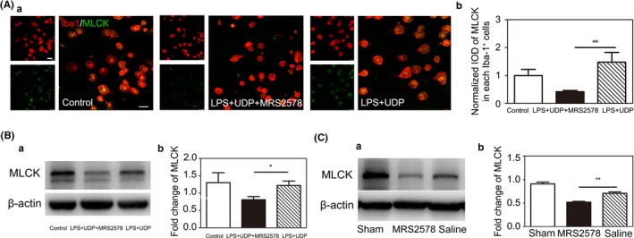
Figure 6.

P2Y6 receptor antagonist MRS2578 inhibit MLCK expression in mice at 3 d after tMCAO and cultured microglia. A, (a) Representative fluorescence images of MLCK in Iba‐1+ cultured primary microglia. Scale bar = 25 μm. (b) Quantitative analysis of MLCK in each Iba1+ microglia. n = 4 per group. B, (a) Western blot for the expression of MLCK in the control, LPS+UDP and LPD+UDP+MRS2578 group. (b) Quantification for the intensity ratios of MLCK/β‐actin in the control, LPS+UDP and LPD+UDP+MRS2578 group. C, (a) Western blot for the expression of MLCK in the sham, saline, and MRS2578 group at 3 d after tMCAO. (b) Quantification for the intensity ratios of MLCK/β‐actin in the sham, saline, and MRS2578 group. Data were presented as mean ± SEM. n = 3‐4 per group. *P < .05, **P < .01
