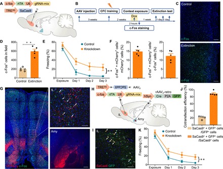
Fig. 5. Perturbing cbp in presynaptic IL extinction-ensemble cells via the CRISPR-SaCas9 system impairs extinction learning.

(A) A schematic of the experiment: A mixture (1:1 ratio) of AAV9–c-fos–rtTA-U6-gRNA-mix and AAV9-TRE3G-SaCas9 was injected into bilateral IL. (B) Scheme of behavioral testing. Rats were trained in the context for fear conditioning 3 weeks after AAV injection. Two weeks later, the rats were given Dox for 1 day, followed by exposure to the context for labeling IL extinction-ensemble cells. Rats were reexposed to the context for three consecutive days for extinction test. (C) Immunofluorescence staining of c-Fos in the IL after context exposure. Scale bars, 200 μm. (D) c-Fos+ cell counts in the IL following context exposure (t test, t8 = 8.18, **P < 0.01; n = 5 slices from three animals in either group). (E) Averaged freezing time during the recall test for cbp knockdown and control groups (two-way ANOVA, **P < 0.01; n = 8 per group. Group effect: F1,28 = 19.45, P < 0.01; time effect: F3,28 = 42.97, P < 0.01; interaction: F3,28 = 0.76, P > 0.05). Rats not exposed to Dox served as control. (F) Graph depicting the percentage of c-Fos+ + mCherry+/mCherry+ (left) and c-Fos+ + mCherry+/c-Fos+ (right) neurons after context exposure (n = 5 slices from three animals). mCherry+: amygdala-projecting IL neurons; c-Fos+: activated neurons. Amygdala-projecting IL neurons (14%) were activated (c-Fos+ + mCherry+/mCherry+), and 12% of the activated neurons projected to amygdala (c-Fos+ + mCherry+/c-Fos+). (G) Illustration of amygdala-projecting IL neurons (mCherry+) that showed immunolabeling of c-Fos protein 2 hours after context exposure. Scale bars, 100 μm (IL) and 200 μm (Amy). (H) Schematic of the experiment: rAAV2-retro-hSyn-Cre-P2A-GFP was injected bilaterally into the amygdala, and a mixture (1:1 ratio) of AAV9–c-fos–rtTA-U6-gRNA-mix and AAV9-TRE3G-DIO-SaCas9 was injected into bilateral IL. (I) Coronal sections showing some amygdala-projecting IL neurons (GFP+) colabeled with Cre- and activity-dependent SaCas9. Scale bar, 100 μm. (J) Cotransfection efficiency of GFP and SaCas9 (n = 5 slices from three animals). Dependent expression of SaCas9 on Cre-GFP resulted in 90% SaCas9+ + GFP+/SaCas9+ neurons. (K) Averaged freezing time during extinction test for cbp knockdown and control groups (two-way ANOVA, **P < 0.01; n = 8 per group. Group effect: F1,28 = 10.64, P < 0.01; time effect: F3,28 = 88.17, P < 0.01; interaction: F3,28 = 0.14, P > 0.05). Rats that were never exposed to Dox served as control.
