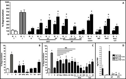
Fig. 2. Activated platelets secrete PAF to stimulate MC degranulation in vitro.

(A) Isolated platelets were activated for indicated periods (5, 15, and 30 min) and at indicated concentrations (1 × 10⁷, 5 × 10⁶, 2.5 × 10⁶, and 1× 10⁶ platelets/ml) with thrombin (or convulxin where indicated). Two MC lines [ROSA (R) and LAD2 (L)] were then exposed to cell-free supernatant from this reaction and MC degranulation measured by tryptase activity assay. In addition, supernatant from activated platelets was ultracentrifuged, and the pellet—resuspended in Tyrode’s buffer (MP, microparticle pellet)—or the supernatant (MPS, microparticle supernatant) was added to MCs. Last, resting platelets (1 × 10⁷) were freeze-thawed and centrifuged, and debris-free supernatant was tested on MCs (Rest F/T). T, Tyrode’s buffer; I, ionomycin positive control; R, resting platelet supernatant. (B) For biochemical characterization of MC-activating effect, LAD2 cells were exposed to supernatant from activated platelets without further treatment (AP), after boiling for 30 min (APb), incubation on activated charcoal (APc), or following isolation of lipid fraction (AP-l). T-l, lipid fraction from Tyrode’s buffer; R-I, lipid fraction from resting platelet supernatant. (C) LAD2 cells were pretreated with antagonists against various lipid mediators [BAY-u 3405 (10 μM): Bay; L798,106 (100 nM): L798; Ex26 (10 μM): Ex; AH 6809 (10 μM): AH; montelukast (100 μM): Mo; WEB2086 (0.1 to 100 μM): Web] before exposure to heat-treated activated-platelet supernatant. Purified PAF was added at 0.1 to 10 μM. Degranulation was measured using β-hexosaminidase assay. NS, not significant. (D) Quantitative determination of PAF in supernatants from resting and activated platelets and activated platelet supernatant absorbed with activated charcoal. Data are represented as the means ± SD. *P < 0.05 versus resting platelet supernatant and #P < 0.05 versus respective activated platelet supernatant, one-way ANOVA and Tukey’s multiple comparisons test. All data derived from four independent experiments were performed in triplicate wells. PRP, platelet-rich plasma.
