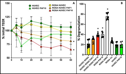
Fig. 3. PAF disrupts endothelial integrity and activates MCs across endothelial barriers.

(A) HUVEC cells were grown to confluency on permeable supports, and then 1 × 10⁶ ROSA cells were added to some of the basal compartments (ROSA HUVEC) followed by addition of PAF at 1 or 10 μM or vehicle control to the apical compartment. TEER was measured for 1 hour. *P < 0.05 versus untreated ROSA/HUVEC cocultures by two-way ANOVA. (B) β-Hexosaminidase from supernatants of HUVEC endothelial cells, ROSA MC cells, and cocultured HUVEC/ROSA cells with or without addition of PAF (at 1 or 10 μM) to the apical side of the endothelia. n = 8 per condition. Results shown as average ± SD, *P < 0.05 versus untreated ROSA/HUVEC cocultures.
