Figure 4.

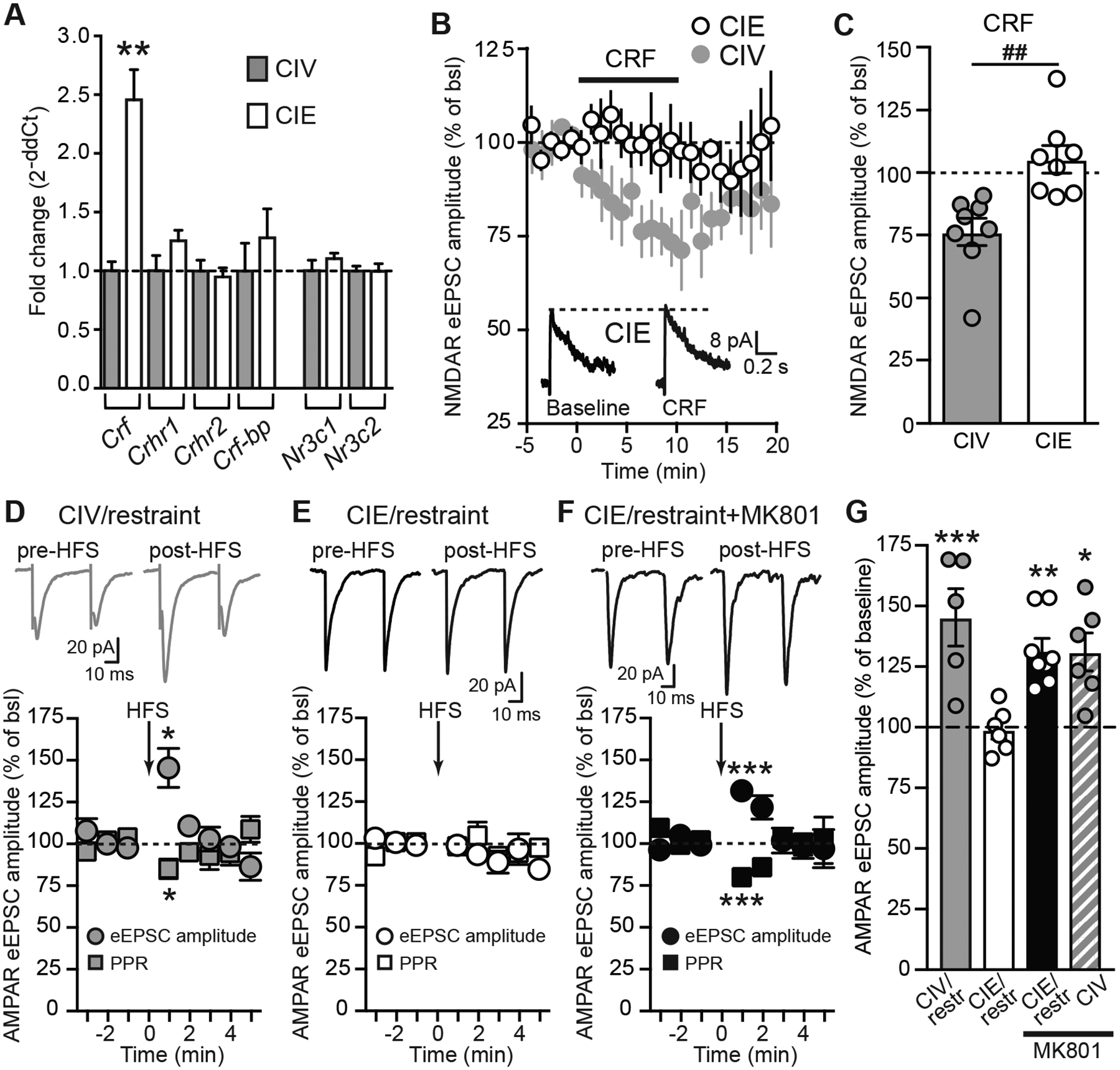
CIE exposure alters CRF expression in the PVN, prevents CRF- and EtOH-induced decreases in NMDAR-currents, and synaptic plasticity of AMPAR-currents in PVN PNCs. A, Summary graph showing the fold change (2−ddCT) of mRNA expression of Crf, Crhr1, Crhr2, Crf-bp, Nr3c1, and Nr3c2 in CIE compared to CIV rats. The fold change in gene expression between CIV and CIE groups was calculated as 2−ddCT (with ddCT=dCT[CIE] − dCT[CIV]). B, Summary time course showing that CIE exposure prevents CRF-induced decreases in NMDAR eEPSC amplitude in PNCs. Summary CIV time course as shown in Fig. 3A. C, Summary graph showing the significant inhibitory effects of CRF in CIV but not in CIE rats. CRF data in bar graph for CIV are duplicate from Fig. 3C. D, Top, representative traces of paired-pulse AMPAR-eEPSCs recorded from PNCs, evoked pre-HFS and post-HFS in slices from CIV/restraint rats. Bottom, summary time course of AMPAR-eEPSC amplitude and PPR pre- and post-HFS. HFS induces a significant increase in eEPSC amplitude and decrease in PPR. EPSC were evoked every 5 sec. The magnitude of STP was calculated as the average amplitude of 12 eEPSCs immediately following HFS (0–1 min post-HFS) and was expressed as % of baseline. E, Top, representative traces of paired-pulse AMPAR-eEPSCs recorded from PNCs, evoked pre-HFS and post-HFS in slices from CIE/restraint rats. Bottom, summary time course of AMPAR-eEPSC amplitude and PPR pre- and post-HFS. HFS-induced STP is impaired in CIE/restraint rats. F, Top, representative traces of paired-pulse AMPAR-eEPSCs recorded from PNCs, evoked pre-HFS and post-HFS in slices from CIE/restraint rats with MK801 (1 mM) included in intrapipette solution. Bottom, NMDAR inhibition with MK801 rescues HFS-induced STP associated with decreased PPR in PNCs from CIE/restraint rats. G, Summary bar graph showing that stress and inhibition of postsynaptic NMDARs unmask synaptic plasticity in CIV rats (CIV/restraint, grey bar; CIV/non-restraint+MK801, grey hatched bar). While CIE exposure prevents stress-induced synaptic plasticity, inhibition of NMDARs rescues STP (CIE/restraint, white bar; CIE/restraint+MK801, black bar).
