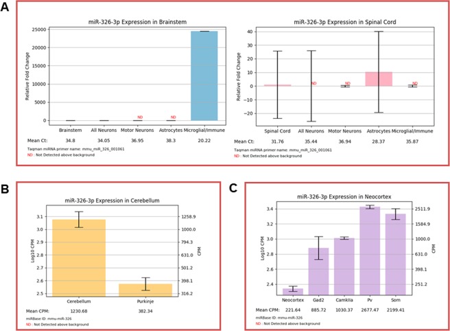
Figure 2.
(A) ‘Search by miRNA’ output for miR-326-3p in the brainstem and spinal cord. Expression is readily detectable exclusively in microglia of the brainstem and astrocytes of the spine (n = 3–4 mice; mean ± SEM). (B) ‘Search by miRNA’ output for miR-326-3p in the cerebellum. miR-326-3p is depleted in Purkinje neurons, relative to the cerebellum as a whole (n = 2–3 mice; mean ± SEM). (C) ‘Search by miRNA’ output for miR-326-3p in the neocortex. miR-326-3p is readily detectable in cortical neurons (n = 2–3 mice; mean ± SEM). Only the more predominant and conserved 3p strand is displayed. The “Valencia” colored frame indicates that miR-326-3p is conserved among most mammals.

