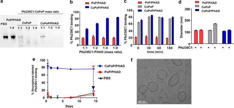
Fig. 1. His-tagged Pfs230C1 spontaneously forms particles when admixed with liposomes containing CoPoP.
a Native PAGE of Pfs230C1 after 1 h incubation with indicated liposomes. The antigen:liposome mass ratio (on CoPoP or PoP basis) is indicated and 1.5 µg of Pfs230C1 was loaded in each lane. The absence of a protein band is indicative of antigen binding to the liposomes, which are too large to migrate in the gel. Representative of three independent experiments. b Binding of Pfs230C1 with varying amounts of CoPoP/PHAD or PoP/PHAD liposomes based on microcentrifugal filtration. c Pfs230C1 binding kinetics based on microcentrifugal filtration. d Size of liposomes before and after Pfs230C1 binding, based on dynamic light scattering. e Serum stability of CoPoP/PHAD with fluorophore-labeled Pfs230C1 during incubation for 2 weeks in 20% human serum at 37 ˚C. The asterisk shows the time after which 0.1% Triton X-100 and proteinase K were added. Graphs show mean ± std. dev. for n = 3 independent experiments. f Cryo EM image of Pfs230C1 with CoPoP/PHAD. Image was taken from a single experiment.

