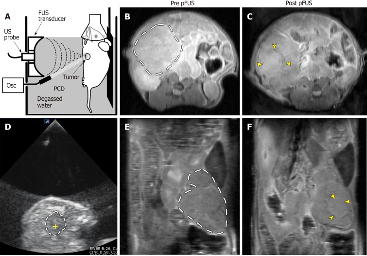
Figure 1.
Representative images of pulsed focused ultrasound treatment and 14T magnetic resonance imaging assessment. (A) Sagittal plane line drawing and (D) axial plane ultrasound (US) image of a pulsed focused ultrasound (pFUS) treatment. Animals were anesthetized, placed on a mobile platform, and partially submerged in degassed water. Tumors were identified using B-mode images from a diagnostic US probe. The KPC mouse tumors generally appear as predominantly hypoechoic masses along the distribution of the pancreas (dashed line in D; the yellow cross within marks the focus of the pFUS transducer). Axial (B and C) and coronal (E and F) pre- and post-treatment proton density weighted anatomic images from a different KPC mouse. Dashed lines in B and E demarcate the pancreatic tumor mass. The treated area demonstrates predominantly isointense signal (solid arrowheads in C and F), with a peripheral ring of hypointense signal (notched arrowheads in C and F), most likely representing sequelae of hyperacute hemorrhage. pFUS: Pulsed focused ultrasound; SC: Subcutaneous; Osc: Oscilloscope; PCD: Passive cavitation detector.

