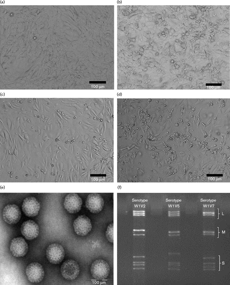
Fig. 1.
Virus culture and identification. (a, c), Uninfected Vero cells (a) and bat cells (c). (b, d), Infected Vero cells (c) and bat cells (d) showed cytopathic changes. (e) Purified viral particles imaged using electron microscopy. (f) Viral genomes observed on polyacrylamide gels. L, M and S indicate the segment group of large, medium and small sizes, respectively.

