Figure 1.

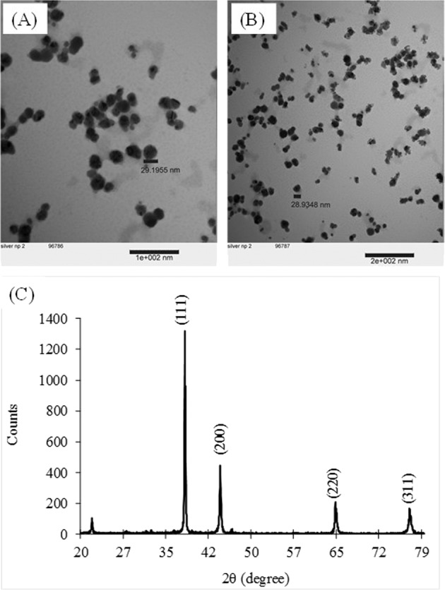
TEM images of silver nanoparticles showing the morphology of synthesized silver nanoparticles at (A) scale bar 100 nm (B) scale bar 200 nm, (C) X-ray diffraction pattern of silver nanoparticles synthesized using onion peel extract.

TEM images of silver nanoparticles showing the morphology of synthesized silver nanoparticles at (A) scale bar 100 nm (B) scale bar 200 nm, (C) X-ray diffraction pattern of silver nanoparticles synthesized using onion peel extract.