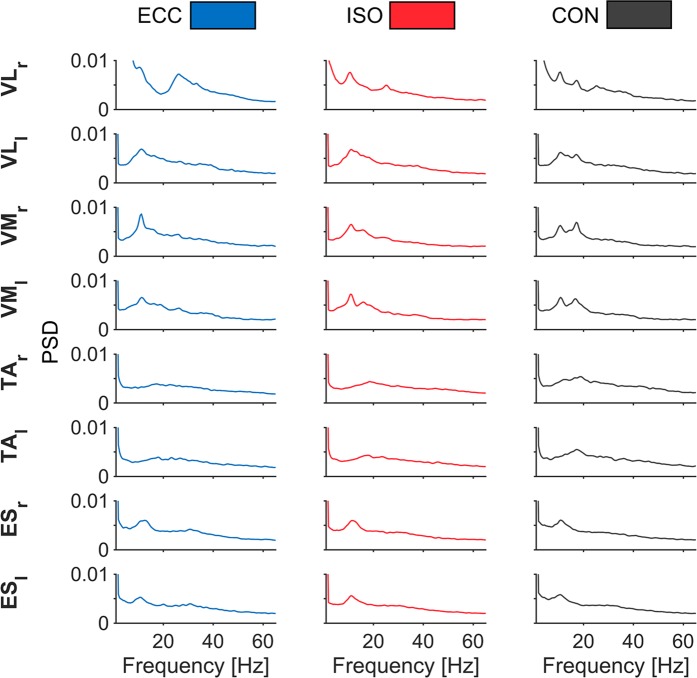
Figure 1.
Normalized PSD of rectified EMG per muscle and period. PSD’s of rectified EMG are depicted for all muscles during each movement period. Power spectra were averaged across muscles, epochs, and participants and normalized to total power. Each column illustrates different movement periods: ECC (blue), ISO (red) and CON (gray). Each row highlights different muscles, with respective labels next to each row. Muscle names are as follows: M. vastus lateralis (VLr & VLl), M. vastus medialis (VMr & VMl), M. tibialis anterior (TAr & TAl), M. erector spinae (ESr, ESl).

