
Figure 4. miR-130a expression causes increased tumor growth in vivo.
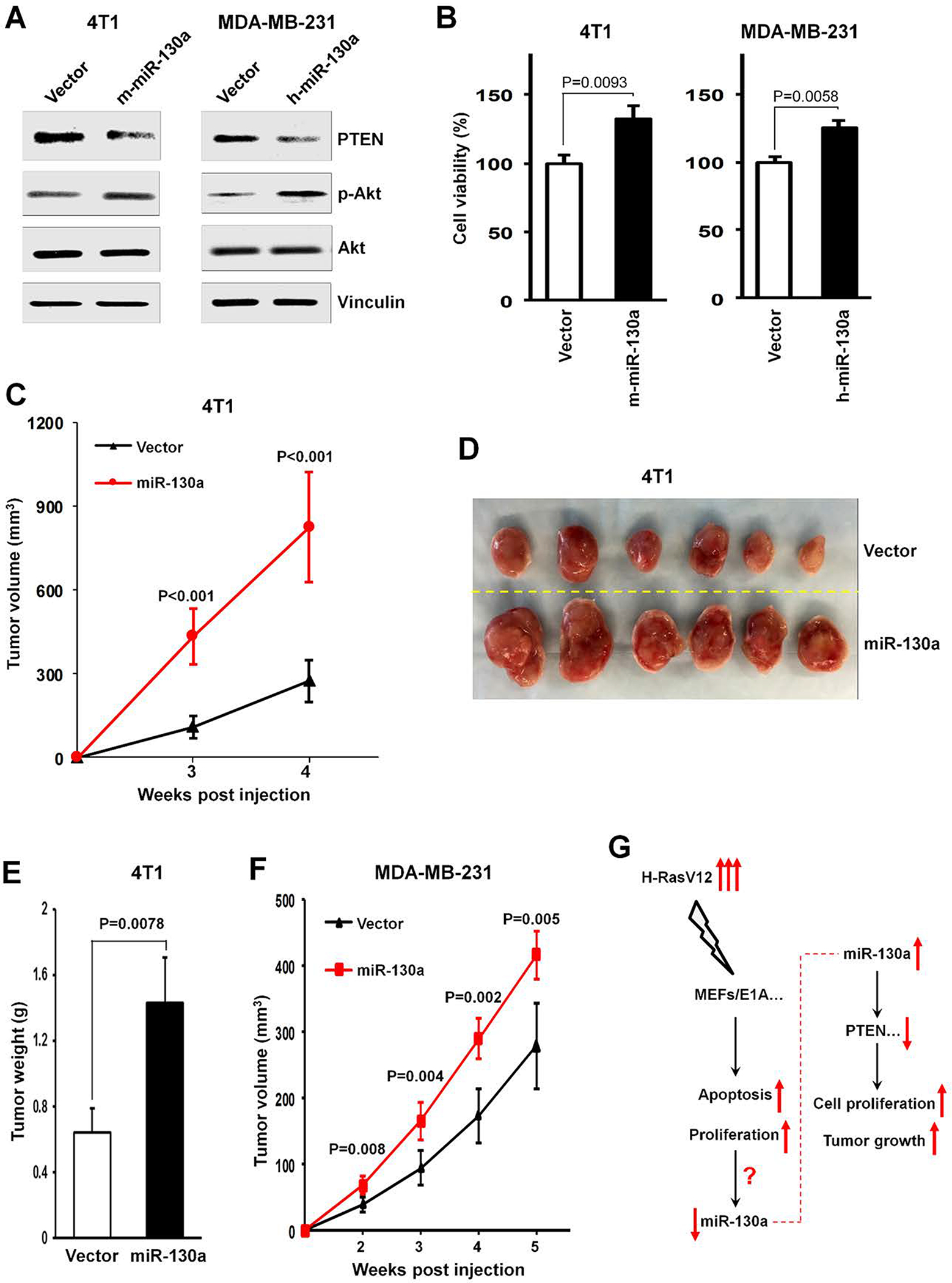
(A, B) 4T1 or MDAMB-231 cells were retrovirally transduced with mouse or human miR-130a expression vectors versus control vector as indicated. Cell lysates were prepared for Western blotting using the indicated antibodies (A). Cell numbers were determined after 5 days of culture. The percent of control cell growth is shown (B). (C) Tumor growth by 5 × 10⁵ 4T1 cells transduced with mouse miR-130a or control vectors. (D, E) Tumor images (D) and tumor weight (E) from mice following subcutaneous injection of 5 × 10⁵ 4T1 cells transduced with mouse miR-130a or control vectors at 4 weeks after implantation. (F) Tumor growth by 5 × 10⁵ subcutaneously injected MDA-MB-231 cells transduced with human miR-130a or control vectors. (G) A schematic outlines the effect of H-RasV12 transient expression in ElA-immorlatalized MEFs (MEFs/E1A) on mediating increased apoptosis and increased proliferation, accompanying by down-regulated miR-130a expression. While oncogenic stress induces a decrease in miR-130a expression, miR-130a triggers increased cell proliferation in vitro and enhanced tumor growth in vivo, by primarily targeting the tumor suppressor, PTEN.