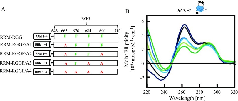
Figure 2.
Effects of Phe in the RGG domain of nucleolin on BCL-2 G-quadruplex folding. (A) Schematic illustration of RRM–RGG and mutated RRM–RGG domains (RRM-RGGF/A1, RRM-RGGF/A2, RRM-RGGF/A3, and RRM-RGGF/A4). (B) Circular dichroism spectra of BCL-2 with RRM-RGG or mutated RRM-RGG domains. Line colors: black, BCL-2; deep blue, BCL-2 and RRM-RGG; blue, BCL-2 and RRM-RGGF/A1; light blue, BCL-2 and RRM-RGGF/A2; yellow green, BCL-2 and RRM-RGGF/A3; and green, BCL-2 and RRM-RGGF/A4. The concentrations of DNA and protein were both 2.5 μM. The G-quadruplex structure is indicated in (B). Black circles in the cartoon of the G-quadruplex represent a guanine residue.

