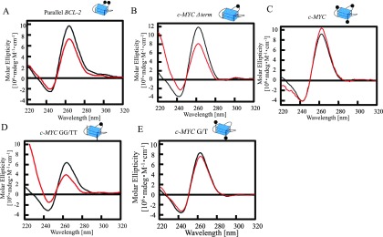
Figure 3.
Properties of RRM in nucleolin for binding to single-stranded DNA and folding several parallel G-quadruplex DNAs. Circular dichroism spectra of Parallel BCL-2 (black) or c- Parallel BCL-2 with RRM (red) (A), c-MYC Δterm (black) or c-MYC Δterm with RRM (red) (B), c-MYC (black) or c-MYC with RRM (red) (C), c-MYC GG/TT (black) or c-MYC GG/TT with RRM (red) (D), and c-MYC G/TT (black) or c-MYC G/T with RRM (E). The concentrations of DNA and protein were both 2.5 μM. The G-quadruplex structure is indicated. Black circles in the cartoons of each G-quadruplex represent a guanine residue.

