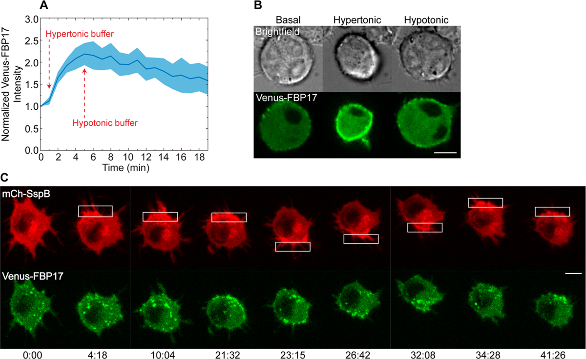
Figure 5.

Changes in plasma membrane tension during protein accumulation-driven migration. (A) Intensity of Venus-FBP17 at the plasma membrane during low and high tension conditions. Cells were transfected with mCh-SspB, iLID-KRasCT, and Venus-FBP17. In the basal state, the cells were incubated with HBSS containing 1 g/L glucose. Hypertonic (low tension) conditions were induced by addition of 1 mL HBSS with 1 g/L glucose and 200 mM sucrose to the dish at t = 1 min. Hypotonic (high tension) conditions were induced by addition of 2 mL H2O to the same dish at t = 5 min. Solid line represents the mean and shaded regions are SEM. n = 3. (B) Representative cell from (A). (C) Distribution of FBP17 during protein accumulation-driven migration. Cell is transfected with mCh-SspB (red), iLID-KRasCT, and Venus-FBP17 (green). FBP17 increased at the photoactivated side in 5 out of 7 cells. Rectangle represents the area of photoactivation. Scale bars are 10 μm. Time is in min:sec.
