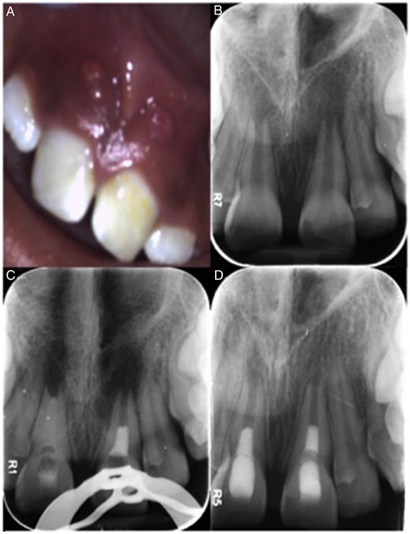
Figure 2.
A 10-year-old female patient with a history of trauma (noncomplicated crown fracture) diagnosed with pulp necrosis and asymptomatic apical periodontitis of teeth #8 and #9. (A) The clinical photograph at the initial visit. Teeth #8 and #9 presented mild swelling and a parulis in the buccal mucosa. (B) Periapical radiolucencies and stage of root formation (Nolla 9) for both apices. (C) The treatment for both was determined by random numbers. Tooth #8 was delayed, and tooth #9 was treated with the immediate induction protocol. (D) At the 12-month follow-up visit, the patient was asymptomatic and showed complete periapical healing and apical healing type 2 with a blunted apex and no evident changes in wall thickening.

