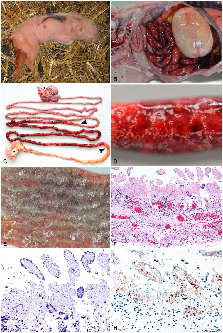
Figure 1.
Typical clinical and pathology presentation of acute necrotic enteritis (NE) in piglets. A. A 1-d-old piglet with hemorrhagic diarrhea. (Photo courtesy H. Nathues, Clinic for Swine, Vetsuisse Faculty, University of Bern.) B. Typical gross lesions of hemorrhagic enteritis in a 1-d-old piglet. C. Typical segmental hemorrhagic and necrotizing jejunitis (between arrowheads). Asterisk = stomach; dot = colon. D. Hemorrhagic intestinal wall with gas bubble formation. E. Mucosa of an acute case of NE. Note that intestinal villi appear white, which is a combination of necrosis and autolysis. Underlying hemorrhage is clearly visible. F. Typical histologic appearance of peracute-to-acute NE in a 1-d-old piglet that died spontaneously. Villi are necrotic and autolytic, with acute hemorrhage in deeper zones of the lamina propria and submucosa. Scant-to-absent inflammatory reaction. (Reprinted with permission.21) 200×. G. Gram stain of the same specimen as panel F depicting necrotic and autolytic villi covered with gram-positive rods. 400×. H. Immunohistochemical detection of Clostridium perfringens beta-toxin (CPB) in the vessels in the lamina propria and submucosa in an acute case of NE. (Reprinted with permission.42) 400×.

