Figure 2.
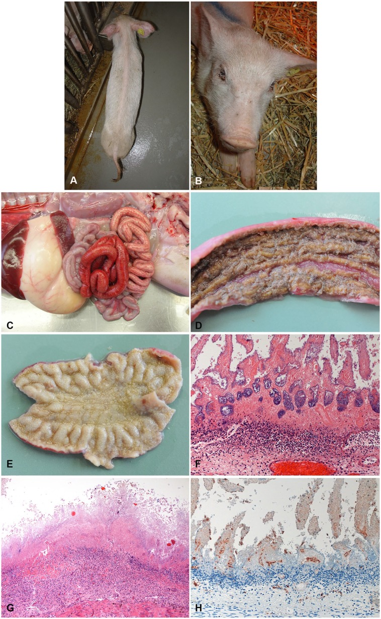
Typical clinical and pathological presentation of subacute-to-chronic necrotic enteritis (NE) in piglets. A, B. External appearance of the same 3-wk-old, severely emaciated (A) and dehydrated (B) piglet. (Photo by N. Wollschläger, courtesy H. Nathues, Clinic for Swine, Vetsuisse Faculty, University of Bern.) C. Typical gross lesions of subacute NE in a 1-wk-old piglet. Segmental hemorrhage (but less severe than in acute case) and fibrin exudation (light red to yellow) in the small intestine. D. Mucosa of a subacute case of NE. Fibrinous pseudomembrane covering the necrotic mucosa. E. Mucosa of a chronic case of NE. Entire mucosa appears yellow because of deep necrosis and inflammatory reaction. F. Typical histologic appearance of the jejunum of a 1-wk-old piglet with subacute NE. Deep necrosis of mucosa and marked, “band-like” neutrophilic infiltration in underlying submucosa. (Reprinted with permission.42) 200×. G. Typical histologic appearance of the jejunum of a 2-wk-old piglet with chronic NE. Complete effacement of the architecture by deep necrosis of the mucosa. Marked inflammatory infiltration in submucosa. 200×. H. Immunohistochemical detection of Clostridium perfringens beta-toxin (CPB) in the vessels in the lamina propria and submucosa in an acute case of NE. 400×.

