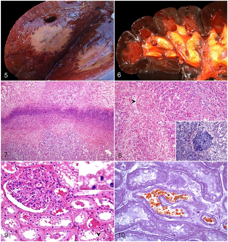
Figures 5–10.
Bovine bacillary hemoglobinuria. Figure 5. Transverse section of liver with a single, large, well-demarcated area of necrosis, and an adjacent acinar pattern. Courtesy of Matias Liboreiro. Figure 6. The renal cortex and medulla are diffusely dark, and the papillary adipose tissue is icteric. Reproduced with permission from Navarro et al. 2016.43 Figure 7. A dense band of leukocytic infiltrate of degenerate neutrophils separates a zone of coagulative necrosis (top) from less severely affected liver (bottom). H&E. Figure 8. Thrombotic occlusion of a central hepatic vein (arrowhead). H&E. Inset: the thrombus is mainly composed of fibrin. Phosphotungstic acid hematoxylin. Figure 9. Renal cortical proximal convoluted tubular epithelium shows variable degrees of cytoplasmic vacuolation and nuclear hyperchromasia. Intracytoplasmic, eosinophilic protein droplets (arrowhead) and luminal casts are present. H&E. Inset: higher magnification of an intracytoplasmic droplet. Figure 10. Intratubular and intracytoplasmic granules shown in Fig. 9 stain orange-brown, consistent with hemoglobin. Okajima stain.

