Figures 11–16.
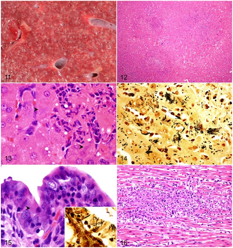
Tyzzer disease. Figure 11. Liver of a foal. Multiple pale necrotic foci distributed throughout the hepatic parenchyma of a foal. Figure 12. Randomly distributed, and variably sized, foci of coagulative necrosis are seen microscopically in the liver. H&E. Figure 13. Intracytoplasmic filamentous bacteria (arrowheads) in hepatocytes at the periphery of a necrotic focus. H&E. Figure 14. Intracytoplasmic bacteria shown in Fig. 13 are better visualized with a silver impression. Steiner stain. Figure 15. The lamina propria of the small intestine of a rabbit is infiltrated and enlarged by numerous mononuclear cells. Filamentous bacteria are observed in the cytoplasm of enterocytes (arrowhead). H&E. Inset: Intracytoplasmic filamentous bacteria in enterocytes are identified with a Steiner stain. Figure 16. Myocardial necrosis, with infiltration by neutrophils, macrophages, and lymphocytes in a rabbit. H&E.

