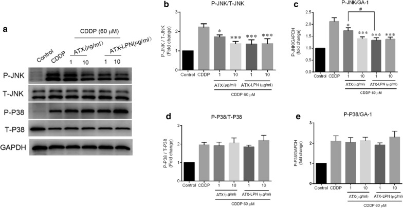
Fig. 6.
Western Blot analyses of the MAPK involvement. a HEI-OC1 cells were treated with CDDP and ATX/ATX-LPN as above. The expression of JNK and p38 were evaluated by WB. b–e Quantification of the light intensity of the protein expression from western blot results above. ATX or ATX-LPN administration on CDDP-treated HEI-OC1 lead to a decreasing ratio of P-JNK/T-JNK (b) and P-JNK/GAPDH (c), with no change in that of P-P38/T-P38 (d) and P-P38/GAPDH (e). *p < 0.05, **p < 0.01, ***p < 0.001 as compared with CDDP. #p < 0.05 as compared with ATX (1 μg/ml),

