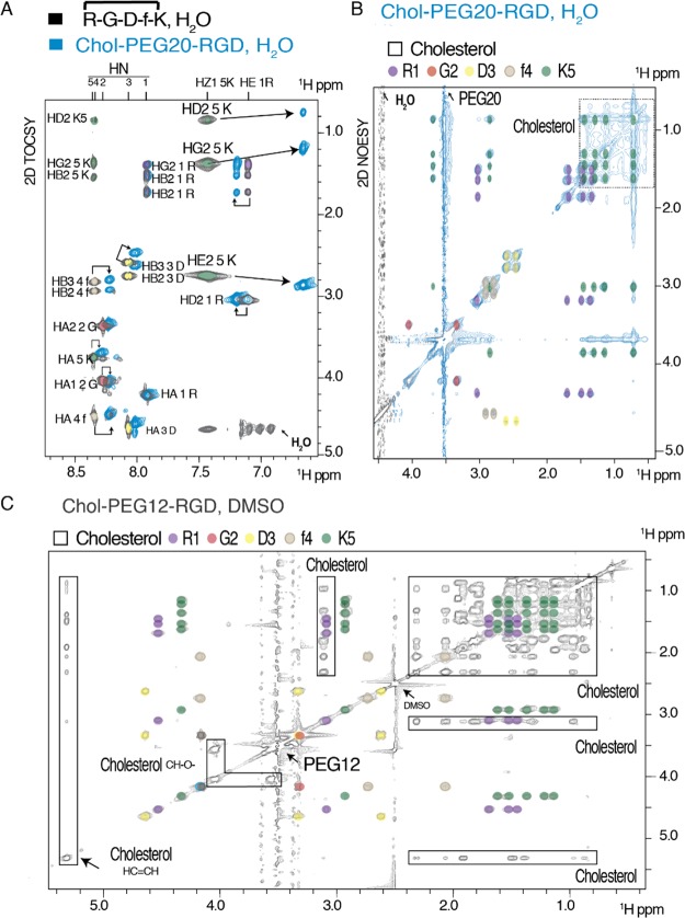
Figure 2.
A: 2D TOCSY (amide-aliphatic region) of the RGD peptide 12 (black) superposed to Chol-PEG20-RGD 17 (blue) in H2O/D2O (9/1). Amino acids are labeled. Chemical shift differences of Lys5 protons upon conjugation to the Chol-PEG20 moiety are labeled and indicated with arrows. B: 2D NOESY of Chol-PEG20-RGD 17 in H2O/D2O (aliphatic region). PEG, cholesterol, and RGD resonances are highlighted. C: 2D NOESY of Chol-PEG12-RGD 15 in DMSO. RGD, cholesterol, DMSO, and PEG12 resonances are indicated.

