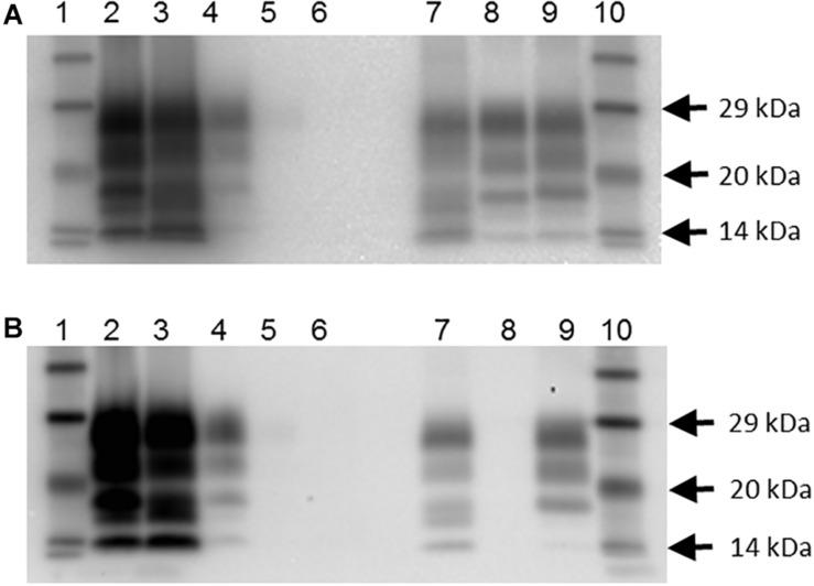
FIGURE 1.
Western blot profile of digested brain samples from selected goats using two monoclonal antibodies. (A) Sha31 antibody. (B) P4 antibody. Lanes 1 and 10: molecular mass marker; lane 2: goat 2135 (clinical suspect, positive on brain by IHC and ELISA); lane 3: goat 2113, clinical suspect, positive on brain by IHC and ELISA); lane 4: goat 2078 (no clinical signs of scrapie, positive on brain by IHC, negative by ELISA); lane 5: goat 2117 (no clinical signs of scrapie, negative on brain by IHC and ELISA, positive on lymphoid tissue by IHC; scrapie profile difficult to discern with the picture contrast used); lane 6: goat 2102 (clinical suspect, negative on brain by ELISA and on all tissues by IHC); lane 7: caprine classical scrapie control (RSCRAP 17/00006, II142QQ222); lane 8: bovine classical BSE control (RBSE 98/00291); lane 9: ovine classical scrapie control (PG1903/97).

