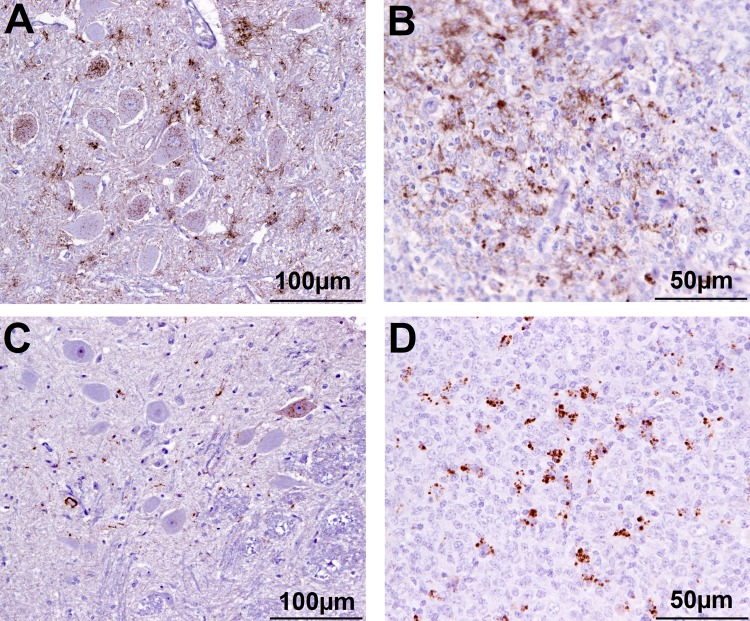
FIGURE 2.
Immunohistochemical examination of brain and lymphoid tissue from selected goats. Obex (A) and medial retropharyngeal LN (B) from clinical suspect goat 2135, positive on brain by ELISA and WB; obex (C) from goat 2146 with inconclusive signs with regard to scrapie, negative on brain by ELISA and WB and pre-scapular LN (D) from clinical suspect goat 2169, negative by all tests on brain, PrPSc detected in pre-scapular LN only. Note the comparatively sparser PrPSc accumulation in the obex in C (immunolabeling visible in only one neuron) compared to A. Immmunolabeling is restricted to tingible body macrophages in the pre-scapular LN in D whereas macrophages and follicular dendritic cells are both immunolabeled in B.

