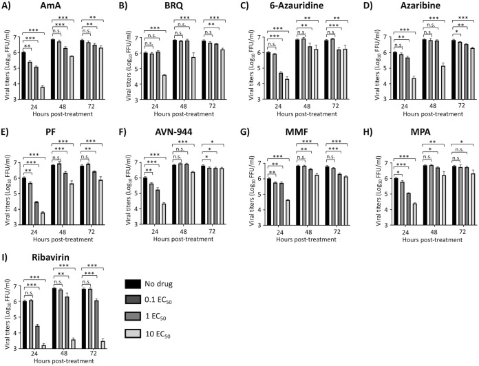
FIG 4.
Inhibition of viral growth kinetics. MDCK cells (24-well plate format; 2.5 × 10⁵ cells/well; triplicates) were infected (MOI = 0.1) with BIRFLU. After 1 h of viral adsorption, infected cells were treated with the indicated 0.1, 1, and 10 EC50 of the different compounds calculated based on the results shown in Fig. 3. Tissue culture supernatants from infected cells were collected at 24, 48, and 72 hpi, and viral titers were calculated using an immunofocus assay (FFU/ml). Data are expressed as mean and SD from three independent experiments conducted in triplicates. Statistical analysis was conducted using an unpaired Student’s t test. *, P < 0.05; **, P < 0.01; ***, P < 0.001; n.s., not significant.

