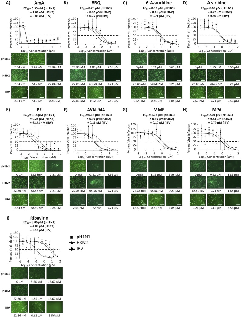
FIG 9.
Inhibition of seasonal H1N1 and H3N2 IAVs and IBV. Confluent monolayers of MDCK cells (96-well plates; 5.0 × 10⁴ cells/well; quadruplicates) were infected with 200 FFU of the indicated Venus-expressing A/California/04/09 H1N1 (pH1N1) and A/Wyoming/3/03 H3N2 IAVs or with B/Brisbane/60/08 IBV. After 1 h of viral adsorption, the indicated concentrations (3-fold serial dilutions, starting concentration of 50 μM) of the different compounds or 0.1% DMSO vehicle control were added to the postinfection medium. Cells treated with 0.1% DMSO vehicle were used as an internal control. At 48 hpi, infected cells were evaluated for viral infection by Venus fluorescence expression using a fluorescence microscope or a fluorescent plate reader. Percent viral infection and the EC50 were calculated based on Nluc expression. Dotted lines indicate 50% viral inhibition. Data are expressed as mean and SD from three independent experiments conducted in quadruplicates. Bar, 50 μm.

