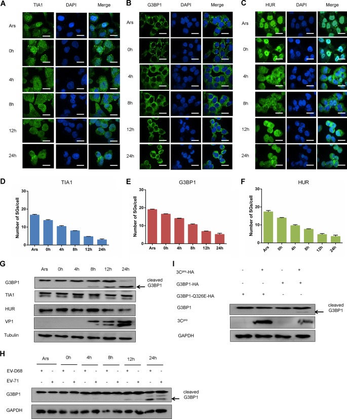
FIG 7.
EV-D68 infection inhibits the formation of SGs responding to Ars. (A, B, and C) RD cells were infected with EV-D68 (MOI of 1), treated with 0.5 μm Ars for 1 h after infection for 0, 4, 8, 12, and 24 h, fixed, and stained with a rabbit anti-TIA1 monoclonal antibody (A), rabbit anti-G3BP1 monoclonal antibody (B), or rabbit anti-HUR monoclonal antibody (C) for immunofluorescence. (D to F) Quantitative analysis of the number of SGs in each cell. (G) RD cells were infected with EV-D68 (MOI of 1). Cells were harvested at 0, 4, 8, 12, and 24 h after infection and treated with Ars for 1 h as a positive control. The influence of EV-D68 on G3BP1, TIA1, and HUR was detected by Western blotting. (H) RD cells were infected with EV-D68 and EV-71. Cells were harvested at 0, 4, 8, 12, and 24 h after infection. Western blotting was used to assess the expression of G3BP1. (I) RD cells were cotransfected with G3BP1 and 3C or G3BP1 Q326E, and 3C. The expression of G3BP1 and G3BP1 Q326E in the cell lysates was detected 48 h after transfection.

