FIG 3.
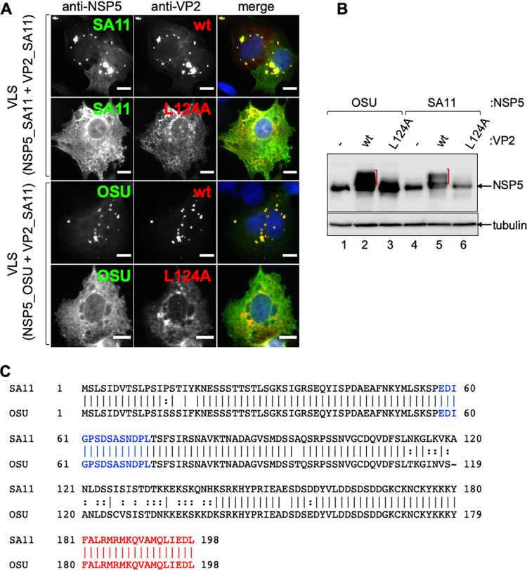
VLS formation and triggering of NSP5 hyperphosphorylation from cognate and noncognate strains are sensitive to VP2 L124A. (A) Immunofluorescence of MA104 cells for detection of VLS composed of NSP5 from simian strain SA11 (top) or NSP5 from porcine strain OSU (bottom) in coexpression with VP2 from simian strain SA11 wt or L124A. At 16 hpt, cells were fixed and immunostained for detection of NSP5 (anti-NSP5, green) and VP2 (anti-VP2, red). A merged image is in the right column of each panel. Nuclei were stained with DAPI (blue). Scale bar is 10 μm. (B) Immunoblotting of lysates from MA104 cells coexpressing NSP5-OSU (lanes 1 to 3) or NSP5-SA11 (lanes 4 to 6) with VP2-SA11 wt (lanes 2 and 5) or L124A (lanes 4 and 6). The membranes were incubated with specific antibodies for the detection of NSP5. Alpha-tubulin was used as a loading control. The red brackets indicate NSP5 hyperphosphorylation. (C) Sequence alignment of NSP5 from simian strain SA11 and porcine strain OSU. The identity between the two proteins corresponds to 94.95%. Noncanonical casein kinase I alpha region and oligomerization tail are labeled in blue and red, respectively.

