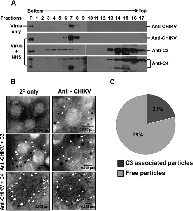
FIG 3.
Exposure of CHIKV to NHS results in deposition of complement components on the virus. (A) Western blot analysis of sucrose density gradient fractions of NHS- or MEM-treated CHIKV. Purified CHIKV was incubated with NHS or MEM and subjected to ultracentrifugation over a preequilibrated 15 to 60% linear sucrose gradient. Fractions of equal volume collected from the bottom of the tube were analyzed for CHIKV antigens and the associated complement components. Complement components C3/C4 comigrated with CHIKV in a linear sucrose gradient when subjected to high-speed ultracentrifugation. Note that fraction 1 is from the bottom, while fraction 17 is the topmost of the gradient as indicated. The migration pattern of viral antigens in both MEM- (top) and NHS-treated CHIKV (2nd from top) was identical to the reactivity observed between fractions 6 to 8. Components of complement proteins C3 (3rd from top) and C4 (bottom) could be seen migrating with CHIKV antigens, but excess or unbound C3/C4 in the CHIKV + NHS sample appeared in the top fractions. P indicates purified CHIKV (top two blots, extreme left lane), C3 (third from the top), and C4 (bottom). The Western blot data comprise a representative image of three independent experiments. (B) Visualization of the C3/C4 component bound to CHIKV by electron microscopy. Chikungunya virus was adsorbed on EM grids and treated with NHS. Bound complement components (C3/C4) to CHIKV were detected after multiple washes followed by probing with anti-human C3 (middle panel, magnification 68,000×) or anti-human C4 (bottom panel, magnification 98,000×) antibody followed by anti-CHIKV antisera and nanogold-conjugated secondary antibodies. The controls included secondary only (top left, magnification 68,000×) and particles probed for viral antigens only (top right, magnification 120,000×). Viral antigens are highlighted by 6-nm (closed triangles) and C3/C4 by 12-nm (arrows) gold secondary antibodies, respectively, with the scale bar indicating 0.025 μm. (C) Pie chart depicting the percentage of particles staining positive for C3 deposition in comparison to C3-negative particles.

