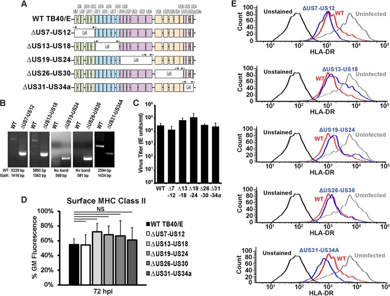
FIG 4.
The unique short region is not required for the downregulation of surface MHC class II molecules in Kasumi-3 cells. (A) Schematic of the US region of the HCMV genome showing the strategy for segmentally knocking out proteins expressed from the US region. Black arrows indicate primer sets either flanking the galK insertion region or binding within galK and a neighboring flanking region. (B) PCR analysis showing replacement of the indicated segment of the US region with galK. The expected values for wild-type and galK-containing bands are indicated below the images. (C) Infectious titers at 120 hpi of wild-type TB40/E (WT) and the US deletion mutants (indicated by Δ7-12, Δ13-18, Δ19-24, Δ26-30, and Δ31-34A) following the infection of fibroblasts at an MOI of 3. (D) Bar graph generated from the geometric mean fluorescence values of surface HLA-DR staining of Kasumi-3 cells infected with wild-type TB40/E or viruses lacking segments of the US region gene (the ΔUS7-US12, ΔUS13-US18, ΔUS19-US24, ΔUS26-US30, and ΔUS31-US34A viruses) at 72 hpi. NS, not significant. (E) Histograms from one representative experiment for the samples for which the results are shown in panel D. The values in panels C and D are averages from three independent experiments.

