FIG 5.
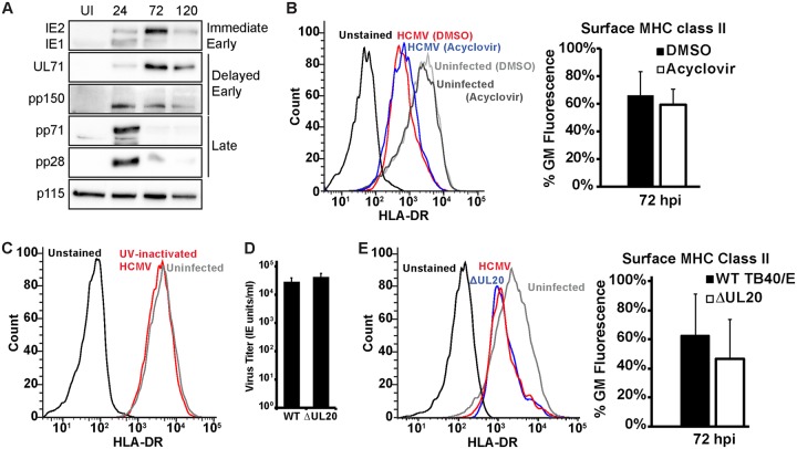
Reduced MHC class II protein requires early viral gene synthesis. (A) Western blot analysis of uninfected (UI) or HCMV-infected Kasumi-3 cells at 24, 72, and 120 hpi, showing the results for two immediate early proteins (IE1 and IE2), two delayed early proteins (UL71 and pp150), two late proteins (pp71 and pp28), and a loading control (p115). (B) (Left) Histograms of one representative experiment of surface HLA-DR staining of uninfected Kasumi-3 cells or cells infected with wild-type TB40/E and treated with DMSO or acyclovir. (Right) The bar graph shows the geometric mean fluorescence values for infected samples at 72 hpi. (C) Histograms of one representative experiment of surface HLA-DR staining at 72 hpi of Kasumi-3 cells infected with UV-inactivated virus. (D) Infectious titers at 120 hpi of wild-type TB40/E (WT) and the ΔUL20 viruses following infection of fibroblasts at an MOI of 3. (E) (Left) Histograms of one representative experiment of surface HLA-DR staining at 72 hpi of Kasumi-3 cells infected with wild-type TB40/E or a virus expressing GFP in place of UL20 (ΔUL20). (Right) Bar graph generated from geometric mean fluorescence values. The values graphed in panels A, D, and E are averages from a minimum of three independent experiments.

