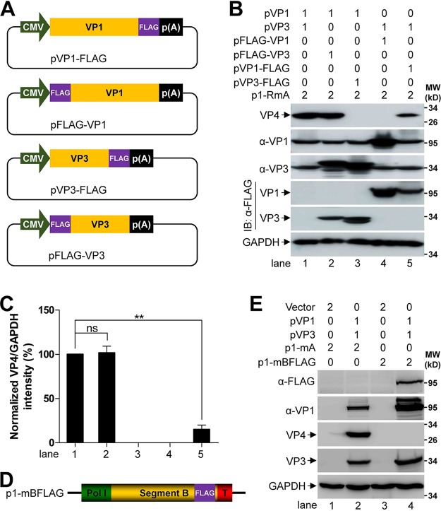
FIG 2.
FLAG tag fused on the C terminus of VP1 reduces IBDV RNP activity. (A) Schematic representation of the plasmids of pVP1-FLAG, pFLAG-VP1, pVP3-FLAG, and pFLAG-VP3. CMV and p(A) stands for cytomegalovirus immediate early promoter and polyadenylation signal, respectively. (B) Monolayer of 293T cells (6-well format) were cotransfected with the indicated plasmids, and the amount of each plasmid is indicated above the lanes (in microgram). Then, the whole-cell lysate was analyzed by Western blot using the antibodies against VP1, VP4, VP3, and FLAG at 72 h posttransfection, and GAPDH was probed as a loading control. (C) Comparison of the expression level of VP4 in different lanes in panel B. The optical density of VP4 and GAPDH was quantified by Quantity One software, and the value of VP4 over GAPDH in the lane 1 of panel B was normalized as 100%. Data were presented as mean ± SD. ns, not significant; **, P < 0.01. (D) Schematic representation of the constitution of p1-mBFLAG. Pol I and T stands for human RNA polymerase I promoter and mouse Pol I terminator, respectively. FLAG tag was fused on the C terminus of VP1. (E) Monolayer of 293T cells (6-well format) were cotransfected with the indicated plasmids, and the amount of each plasmid is indicated above the lanes (in microgram). Then, the whole-cell lysate was analyzed by Western blot using the antibodies against VP1, VP4, VP3, and FLAG at 72 h posttransfection, and GAPDH was probed as a loading control.

