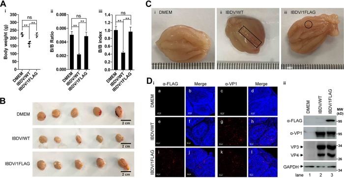
FIG 4.
Attenuation of IBDV pathogenicity by fusing FLAG tag on the C terminus of VP1. (A–C). One-week-old SPF chickens (5 in each group) were inoculated with DMEM (0.2 ml each), IBDV/WT (104 PFU each), or IBDV/1FLAG (104 PFU each) through eye dropping. After 14 days of regular isolated feeding, the bodyweight was determined before sacrifice (Ai), the BFs was isolated and weighed and the weight ratio of BFs to bodyweight (B/B ratio) was calculated (Aii), and the B/B index was determined (Aiii) according to the method described in the section of Materials and Methods; at the same time, the images of the overall BFs appearance (B) and the interior of representative BFs of chickens inoculated with DMEM (Ci), IBDV/WT (Cii), or IBDV/1FLAG (Ciii) were photographed, and the hemorrhagic sites or the single hemorrhagic points were squared or circled in the BFs interior representative images. Scale bar in panel B stands for 2 cm, and the distance between two adjacent lines in panel C is 1 mm. Data were presented as mean ± SD. ns, not significant; **, P < 0.01. (D) The BFs sections of chickens inoculated with DMEM, IBDV/WT, or IBDV/1FLAG were immunostained with the antibodies against FLAG (frames a, e, and i) or VP1 (frames c, g, and k) followed by staining with Alexa Fluor-568-labeled secondary antibody, and the nucleus was stained with DAPI (panel i). Scale bars = 50 μm. The whole lysate of the BFs, which were from the chickens inoculated with DMEM, IBDV/WT, or IBDV/1FLAG, was also subjected to Western blot analysis by using the antibodies against FLAG, VP1, VP3, and VP4, and GAPDH was probed as a loading control (panel ii).

