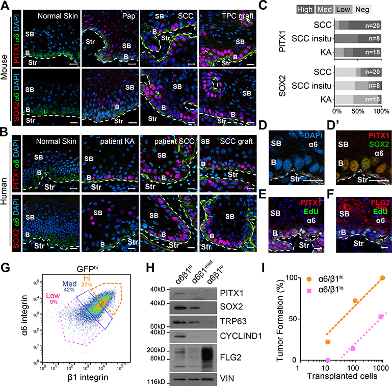
Figure 1: PITX1 is de novo expressed in basal tumor propagating SCC cells.
(A,B) Confocal micrographs of mouse (A) and human (B) tissue sections. Basal (b), supra-basal (sb), and stromal (Str) cells in papillomas (Pap), primary SCCs (SCC) from mice and patients, patient keratoacanthoma (KA), and TPC grafts. Scale bars = 50μm. (C) Stacked bar graphs illustrate the prevalence of PITX1 and SOX2 expression in n patient specimens. (D-F) Confocal microscopy shows PITX1 colocalizes with SOX2 (D’) and EdU (E,F) in basal (b) but not Filaggrin (F) expressing supra-basal (sb) SCC cells. Scale bars = 50μm. (G) FACS separation of α6- and β1-integrin (CD49f and CD29) high (Hi), medium (Med) and low (lo) fractions of GFP marked SCC parenchyma. (H) Western blotting of parenchymal fractions with SOX2, PITX1, TRP63, and CYCLIN D1, and FLG2 antibodies. (I) Limited-dilution transplantation experiments of SCC fractions. (n=18 transplants; 3 independent SCCs). See also Figure S1.

