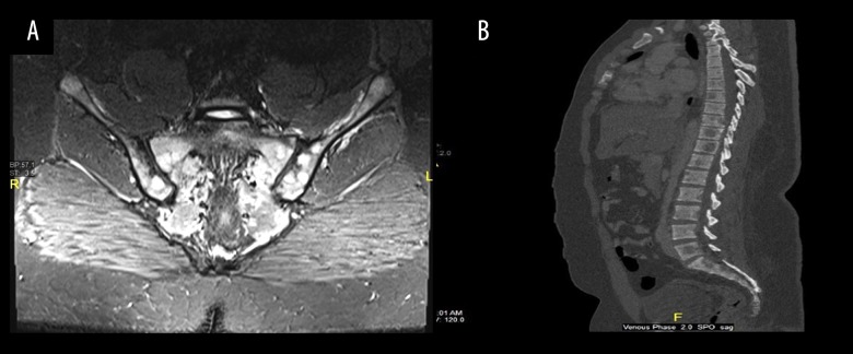
Figure 2.
Magnetic resonance imaging (MRI). (A) Coronal SITR of the sacrum and sacroiliac joints showing multiple bright foci of infiltration are noted bilaterally involving the sacrum and iliac side of both sacroiliac joints. (B) Sagittal reconstruction of the dorso-lumbosacral spine (bone window setting) showing multiple hypodense lytic lesions notably at L5, D11, D10, D9 as well as the proximal sacral pieces.

