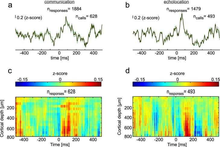
Fig 2. LFPs during vocalization in the CN and FAF.
(a) Mean LFP (± SEM) of all isolated communication calls (n = 628) studied. Signals from all three channels of the striatum were pooled together thus rendering a higher number of responses for the striatum than for the FAF. (b) Mean LFP (± SEM) obtained during the production of isolated echolocation pulses (n = 493) in the striatum. (c) and (d) Colormaps showing the mean of z-scored LFPs in the FAF across cortical depths, 500 ms before and after communication calls (c) and echolocation pulses (d). Data underlying this figure can be found at https://doi.org/10.12751/g-node.6a0d94. CN, caudate nucleus; FAF, frontal auditory field; LFP, local field potential.

