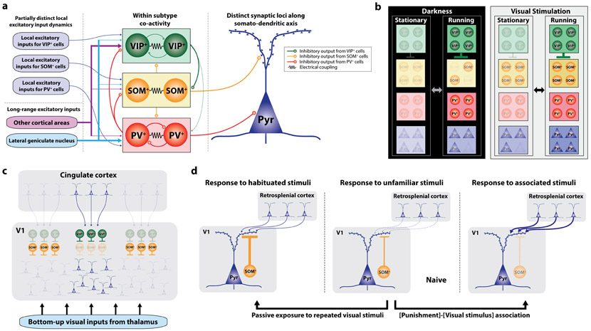
Figure 1: Context-dependent modulation of V1 by IN subtypes.
(a) A wiring diagram illustrating differential excitatory inputs, local inhibitory connectivity and inhibitory outputs in V14-7,23. (b) Locomotion-dependent modulation of IN activity in the presence or absence of concurrent visual inputs16,18. In the darkness, locomotion activates both PV-INs and VIP-INs, but SOM-INs and pyramidal neurons are heterogeneously modulated. With visual stimuli, locomotion activates all IN subtypes and pyramidal neurons. Color of each cell type reflects the activity level relative to its level during stationary state in darkness. (c) Top-down projections from cingulate cortex to specific retinotopic site in V1 induce local disinhibition by preferentially recruiting VIP-INs. In contrast, SOM-INs increase their activity at surrounding areas and cause surround suppression. To our knowledge, it has not been determined whether the cingulate neurons projecting to different V1 locations are intermingled or topographically organized as in the frontal eye field that mediates attentional modulation in primate visual cortex28. (d) Context-dependent gating of top-down inputs from retrosplenial cortex by SOM-INs37. Visual responses in SOM-INs increase after passive exposure to repeated visual stimuli. In contrast, visual responses in SOM-INs decrease when the mouse learns association between the visual stimulus and tail shock, permitting strong top-down modulation of visual response in excitatory neurons by retrosplenial cortex. Line thickness and opacity of cell reflect the connectivity strength and the activity, respectively.

