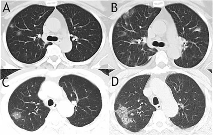
Fig 4. Two cases of CT images from survival group showed ground glass nodular opacities on admission and progressed to multiple patchy ground glass opacities on reexamination.
The CT images of a 60-year-old woman on admission showed peripherally distributed focal ground glass nodular opacities in only right lung (A) and rechecked CT images showed expanded area of patchy ground glass opacities in both right and left lungs with reticular and interlobular septal thickening on 7 days later (B). Unilateral ground glass nodular opacity was found on CT images of a 64-year-old man from survival group (C) and progressed patchy ground glass opacities as well as interlobular septal thickening were seen after 4 days (D).

