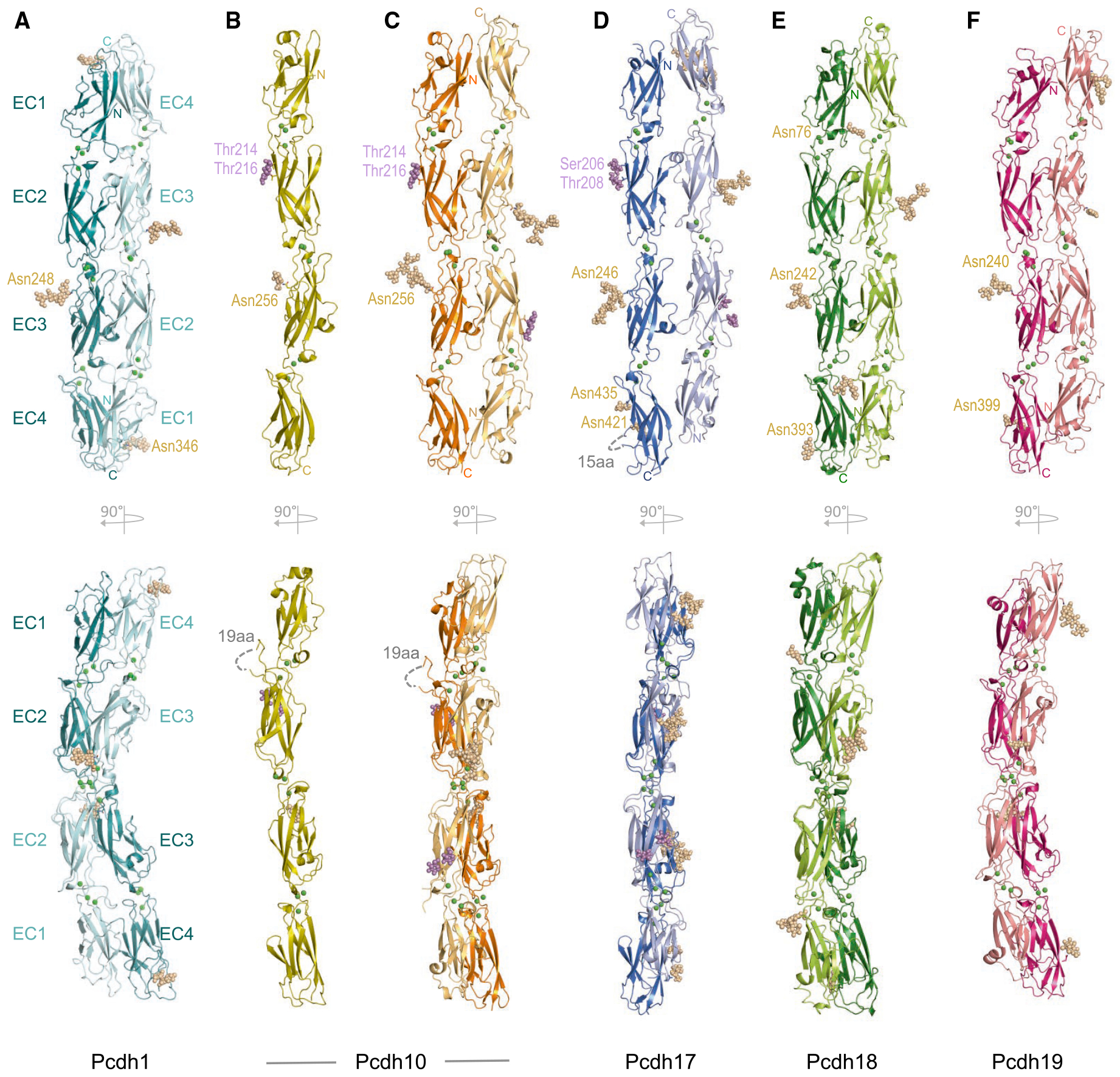
Figure 2. Structures of Adhesive EC1–EC4 Fragments of δ1- and δ2-Protocadherins.

(A–F) Ribbon representations showing two orthogonal views (upper and lower panels) of human EC1–EC4 fragment structures of (A) pcdh1; (B) pcdh10 monomer; (C) pcdh10 dimer; (D) pcdh17; (E) pcdh18.; and (F) pcdh19.
Single trans dimers, formed between symmetry-related protomers (A) or in the crystallographic asymmetric unit (C–F) are shown. Interdomain calcium ions are shown as green spheres; N-linked and O-linked glycans as wheat and magenta spheres. See Figures S3 and S4 and Tables S1 and S2.
