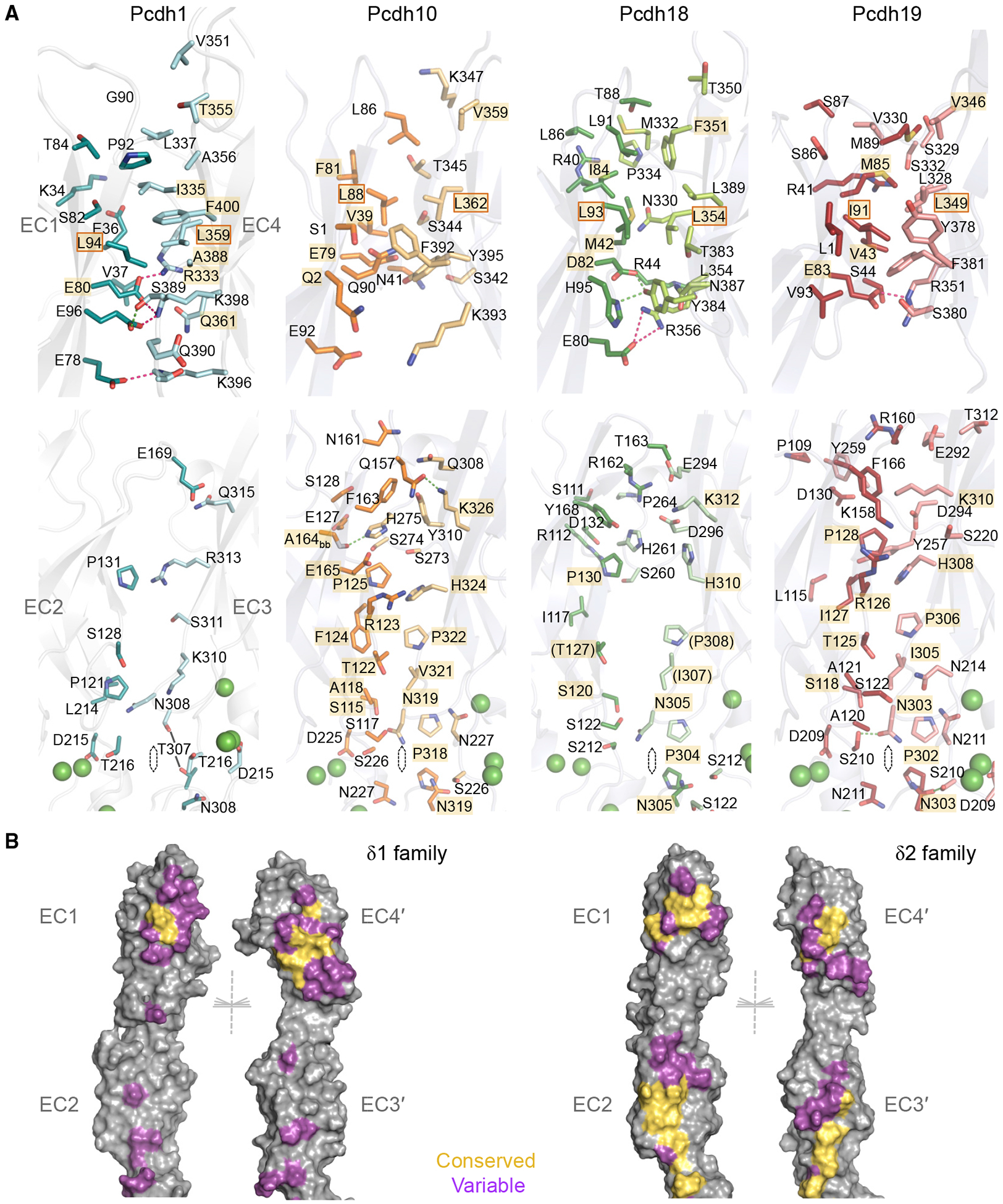
Figure 3. Conserved and Variable Molecular Interactions in the trans Dimer Interface.

(A) Residue views of interface regions EC1:EC4 (top) and EC2:EC3 (bottom) in trans dimers of pcdh-1, −10, −18, and −19 (left to right). Side chains of interfacial residues (>5% buried) are shown as sticks. Interactions conserved between multiple structures are highlighted in gold. Conserved hydrophobic residues chosen for mutation are boxed. Green spheres: calcium ions.
(B) Molecular surfaces of representative δ1 (pcdh1, left) and δ2 (pcdh10, right) trans dimer structures opened to display interfacial residues color-coded according to their conservation within the respective subfamily (yellow: conserved in character; magenta: variable). Non-interface residues are shown in gray. Half of each 2-fold symmetric dimer is shown for clarity.
See Figures S3–S5.
