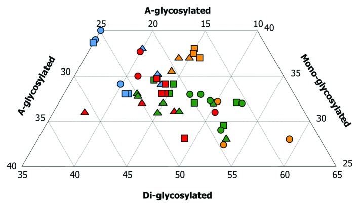
Figure 4. Triplot analyses from brains of sheep infected with cloned murine strains. Colors denote strain whereas symbol denotes breed and genotype. This plot shows total number of animals infected either by a single oral dose or a combined challenge. Eighteen sheep infected with ME7 (green) gave a positive result in Western-blot. Nine did with 79A (blue). Eleven sheep infected with 22A (red) gave a positive result in WB. All nine sheep infected with 87V (yellow) displayed strong signals of PrPres in their brains. Note that the PrPres associated with ovine ME7 showed higher amounts of the di-glycosylated band, followed by intermediate amounts of the mono-glycosylated band and low amounts of the a-glycosylated band. The characteristics of PrPres in ovine 22A infections are similar to ME7 ones but with a relatively lower and higher amount of the di- and a-glycosylated bands, respectively. The mono- and the a-glycosylated bands of PrPres are significantly higher after 79A infections. This pattern was similar to that observed for the PrPres associated with the 87V strain in Cheviot sheep of the VRQ/VRQ (triangles) and ARQ/ARQ (squares) genotypes, but not in the Suffolk sheep of the ARQ/ARQ genotype (circles), in which, the di-glycosylated band predominated.

An official website of the United States government
Here's how you know
Official websites use .gov
A
.gov website belongs to an official
government organization in the United States.
Secure .gov websites use HTTPS
A lock (
) or https:// means you've safely
connected to the .gov website. Share sensitive
information only on official, secure websites.
