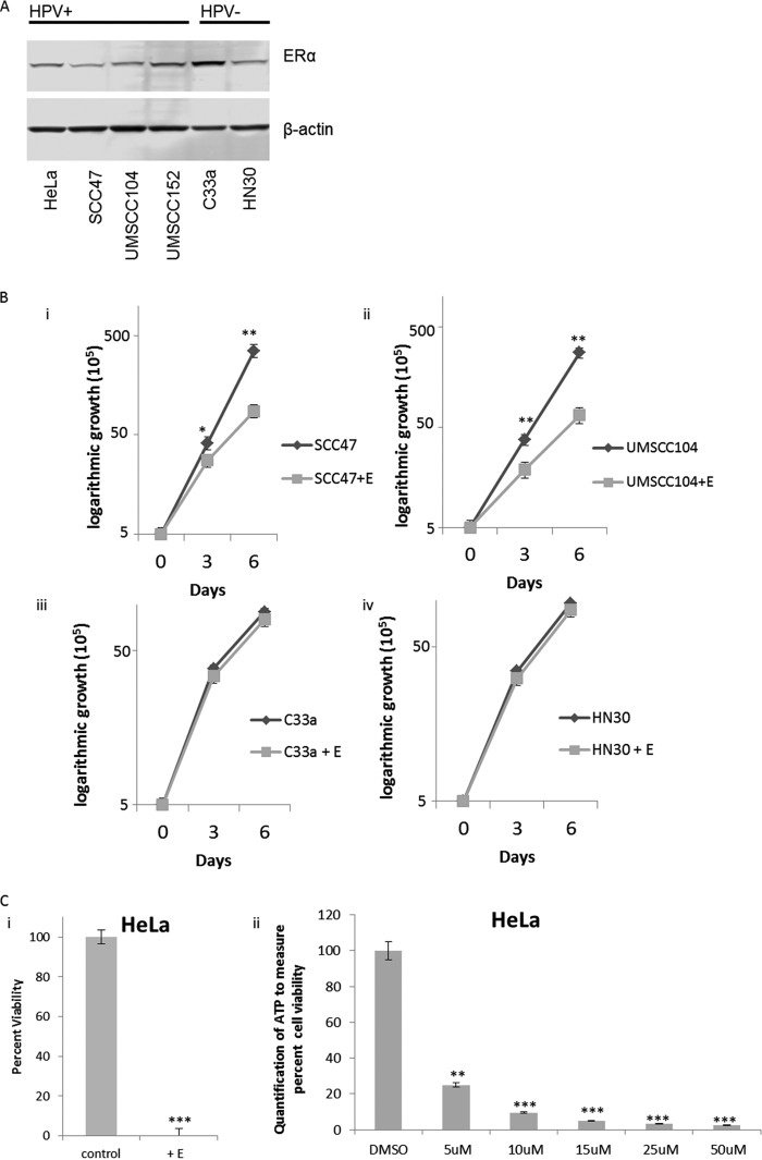
FIG 1.
Estrogen attenuates the growth of HPV-positive (HPV+) or HPV-negative (HPV−) cancer cell lines. (A) Cervical cancer cell lines HeLa and C33a, as well as HNSCC cell lines SCC47, UMSCC104, UMSCC152, and HN30 were analyzed for their expression of the ERα and compared to the loading control β-actin. HPV status is indicated above the blots. Experiments were conducted in triplicate, and no significant correlation between HPV status and ERα expression was observed. (B) HPV+ SCC47 (i) and UMSCC104 (ii) cells and HPV− C33a (iii) and HN30 (iv) cells were seeded on day zero and grown in the presence of 15 μM estrogen (+E) or absence of estrogen. Cells were trypsinized and counted on days 3 and 6, and cell counts are presented on a logarithmic scale. Statistical differences in both SCC47 and UMSCC104 cells can be observed at both days 3 and 6. Values that are significantly different are indicated by asterisks as follows: *, P < 0.05; **, P < 0.001. No statistical difference is observed between treatments on day 3 or day 6 in C33a cells (iii) or HN30 cells (iv). Experiments were conducted in triplicate, and error bars are representative of the standard errors (SE). (C) (i) HeLa cells were grown in the presence or absence of 15 μM estrogen for 72 h, and then cells were counted for viability via trypan blue exclusion. (ii) Data are presented as percent viability at 48 h as measured by luciferase to monitor ATP via the Promega Cell Titer-Glo assay, over DMSO control. Experiments were conducted in triplicate, and error bars are representative of SE. **, P < 0.001; **, P < 0.001.

