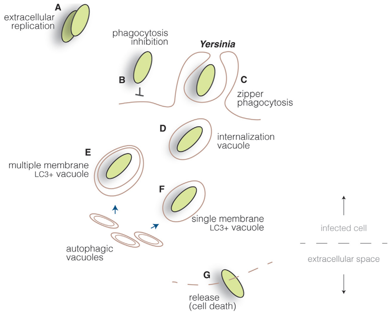
Fig. 4.
Yersinia cycle. Yersinia proliferate mainly as extracellular pathogens (A), as they actively inhibit cellular uptake by injecting type III secretion system effectors into host cells, blocking actin cytoskeleton rearrangements (B). However, through the interaction of surface invasion proteins and host cell receptors, Yersinia can induce its internalization in specific cell lines (C). Bacteria are found initially within internalization vacuoles (D) which, through interaction with autophagic vacuoles, can mature into multiple (E) or single (F) membrane compartments, depending on the cell type. Yersinia are probably released upon cell death in cells permissive for bacterial replication (G).

