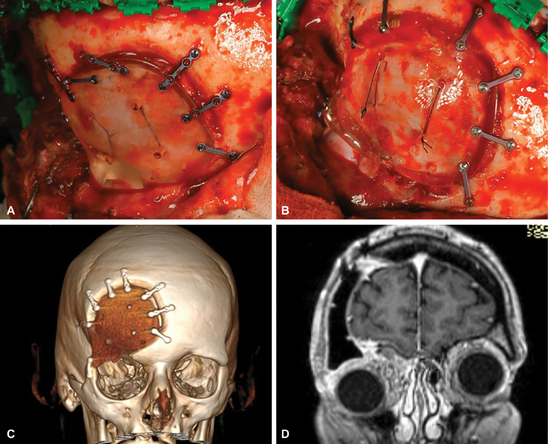
Fig. 4.

A 54-year-old female patient presented with right sided exophthalmos. CT and MRI showed an enplaque meningioma of the sphenoid wing and temporal fossa extending to the orbital rim (data not available). A single-step procedure with subtotal tumor resection, orbital decompression, and acryl CAD/CAM implant reconstruction was performed. Intraoperative photographs show a suboptimal fit of the implant after craniectomy with the drilling template ( A ) with an elevation of the orbital rim ( B ). After the swelling subsided, the patient presented with a persistent depression of the right orbit leading to a facial dysmorphia. Postoperative three-dimensional CT reconstruction revealed suboptimal placement of the implant, with a gap between frontal bone, implant and low lying orbital rim ( C ). Postoperative coronal MRI showing excessive pressure of the contents of the right orbit through the misaligned implant ( D ). Repositioning of the implant in a second procedure was necessary to correct the orbital congestion. CT, computed tomography; MRI, magnetic resonance imaging.
